Loculated Pleural Effusion Ct - Pleural Effusion Pacs Suche Fur Radiologen / The area of increased attenuation (black ∗) in all three images represents collapsed lung (tif 593 kb) fig.
byAdmin-
0
Loculated Pleural Effusion Ct - Pleural Effusion Pacs Suche Fur Radiologen / The area of increased attenuation (black ∗) in all three images represents collapsed lung (tif 593 kb) fig.. This type of effusion is empyema unless proven otherwise. Other causes are complicated parapneumonic effusion , empyema, and tuberculosis. We report the successful treatment of a loculated pleural effusion using intrapleural urokinase in a patient with severe chronic obstructive pulmonary disease. Can demonstrate small effusions (10 ml of fluid or less). The authors develop a method to accurately and easily estimate the volume of pleural effusions with computed tomography (ct).
Ct during catheter placement shows catheter within the loculated collection. Loculated right pleural effusion with foci of atelectasis and consolidative changes concerning for pneumonia. Surgical thoracostomy tube placement and radiologically guided catheter drainage are standard therapy for loculated pleural fluid collections. Empyema is defined by purulent fluid collection in the pleural space, which is most commonly caused by pneumonia. E7.4 ct of pleural effusion.
Pleural Effusion from www.stritch.luc.edu Can demonstrate small effusions (10 ml of fluid or less). Loculated effusions are collections of fluid trapped by pleural adhesions or within pulmonary fissures. Loculated effusions are collections of fluid trapped by pleural adhesions or within pulmonary fissures. Can demonstrate complex, loculated collections and pleural thickening. What are the different appearances of pleural effusion? Bilateral, left greater than right, pleural effusions with adjacent atelectasis and collapse versus consolidation of the left lower lobe. Loculated pleural effusion ct : Download as powerpoint open in image viewer figure 4.
Pleural effusion is an accumulation of fluid in the pleural space that is classified as transudate or exudate according to its composition and underlying pathophysiology.
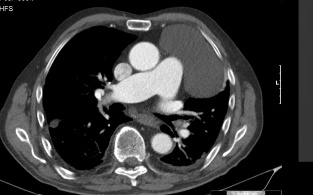
Department of medicine , 250 east supperior st, rm 456, chicago 60611. Other causes are complicated parapneumonic effusion , empyema, and tuberculosis. However, ct can help distinguish between a pleural effusion and a pleural empyema (see pleural effusion vs pleural empyema). Most effusions start like this and can be easily missed. The largest pocket of fluid is present posteriorly at the right lung base, with associated atelectasis and minor consolidation. The authors develop a method to accurately and easily estimate the volume of pleural effusions with computed tomography (ct). In the past, the finding of pleural thickening at ct in patients with pneumonia or neoplasm was found to be highly indicative for the presence of an exudate 6 . Prior chest radiographs indicating that the blunting is a new finding also provide a good indicator of pleural effusion. We report the successful treatment of a loculated pleural effusion using intrapleural urokinase in a patient with severe chronic obstructive pulmonary disease. We studied the value of transca … Posteroanterior and lateral chest radiographs usually confirm the presence of a pleural effusion, but if doubt exists, ultrasound or computed tomography (ct) scans are definitive for detecting. Frequently suggested by the radiologists to image the underlying lung. Loculated right pleural effusion with foci of atelectasis and consolidative changes concerning for pneumonia.
(a) axial, (b) coronal, and (c) sagittal images show a large amount of uniform material of fluid attenuation filling much of the right hemithorax (white ∗). Treatment may fail if the catheter is not placed optimally within the loculation or if the fluid is hemorrhagic or fibrinous. More than one half of these massive pleural effusions are caused by malignancy; Prior chest radiographs indicating that the blunting is a new finding also provide a good indicator of pleural effusion. Most effusions start like this and can be easily missed.
Epos Trade from epos.myesr.org The imaging of pleural effusions will be presented here. More than one half of these massive pleural effusions are caused by malignancy; (a) axial, (b) coronal, and (c) sagittal images show a large amount of uniform material of fluid attenuation filling much of the right hemithorax (white ∗). Ct during catheter placement shows catheter within the loculated collection. Surgical thoracostomy tube placement and radiologically guided catheter drainage are standard therapy for loculated pleural fluid collections. Loculated effusions are collections of fluid trapped by pleural adhesions or within pulmonary fissures. Assesses the pleura for thickening and mass, the chest. 7) chest radiographs show a left posterolateral loculated effusion.
Loculated pleural effusion ct :
In the past, the finding of pleural thickening at ct in patients with pneumonia or neoplasm was found to be highly indicative for the presence of an exudate 6 . Assesses the pleura for thickening and mass, the chest. Treatment may fail if the catheter is not placed optimally within the loculation or if the fluid is hemorrhagic or fibrinous. Loculated pleural effusion (427895005) recent clinical studies. Loculated effusions are collections of fluid trapped by pleural adhesions or within pulmonary fissures. Surgical thoracostomy tube placement and radiologically guided catheter drainage are standard therapy for loculated pleural fluid collections. Pleural effusions are characterized on ct by attenuation values between those of water (0 hounsfield units hu) and soft tissue (approximately 100 hu), typically in the order of 10 to 20 hu. (a) axial, (b) coronal, and (c) sagittal images show a large amount of uniform material of fluid attenuation filling much of the right hemithorax (white ∗). Computed tomography (ct) of the chest is often used (1) and. Ct during catheter placement shows catheter within the loculated collection. All patients require medical management with antibiotics. (b) sagittal reconstruction (parenchymal window). The largest pocket of fluid is present posteriorly at the right lung base, with associated atelectasis and minor consolidation.
Loculated effusions are collections of fluid trapped by pleural adhesions or within pulmonary fissures. Pleural effusion is an accumulation of fluid in the pleural space that is classified as transudate or exudate according to its composition and underlying pathophysiology. (a) axial plane (soft tissue window). However, ct can help distinguish between a pleural effusion and a pleural empyema (see pleural effusion vs pleural empyema). Loculated right pleural effusion with foci of atelectasis and consolidative changes concerning for pneumonia.
Loculated Pleural Effusion Radiology Case Radiopaedia Org from prod-images-static.radiopaedia.org The authors develop a method to accurately and easily estimate the volume of pleural effusions with computed tomography (ct). Download as powerpoint open in image viewer figure 4. The area of increased attenuation (black ∗) in all three images represents collapsed lung (tif 593 kb) fig. Ct during catheter placement shows catheter within the loculated collection. The largest pocket of fluid is present posteriorly at the right lung base, with associated atelectasis and minor consolidation. This type of effusion is empyema unless proven otherwise. Computed tomography (ct) of the chest is often used (1) and. However, ct can help distinguish between a pleural effusion and a pleural empyema (see pleural effusion vs pleural empyema).
The imaging of pleural effusions will be presented here.
7) chest radiographs show a left posterolateral loculated effusion. However, ct can help distinguish between a pleural effusion and a pleural empyema (see pleural effusion vs pleural empyema). Frequently suggested by the radiologists to image the underlying lung. Can demonstrate small effusions (10 ml of fluid or less). What are the different appearances of pleural effusion? Treatment may fail if the catheter is not placed optimally within the loculation or if the fluid is hemorrhagic or fibrinous. A sample of extracted pleural fluid, which is purulent in appearance. Other causes are complicated parapneumonic effusion , empyema, and tuberculosis. (a) axial, (b) coronal, and (c) sagittal images show a large amount of uniform material of fluid attenuation filling much of the right hemithorax (white ∗). More than one half of these massive pleural effusions are caused by malignancy; Can demonstrate complex, loculated collections and pleural thickening. Pleural effusion is an accumulation of fluid in the pleural space that is classified as transudate or exudate according to its composition and underlying pathophysiology. Most effusions start like this and can be easily missed.